
Case 2 – 2024
Learning Outcomes
- To recognise the features of manifest primary open angle glaucoma,
- To understand the basic and advanced principles of Visual field testing.
- To understand and interpret OCT in the context of glaucoma
- To understand the artifacts associated with visual field and OCT
Case:
A 59 year-old Caucasian woman presented for a routine optometry examination. She is emmetropic but has developed presbyopia. She has been wearing over-the-counter readers until now.
She is not aware of a family history for glaucoma. She reports good general health, and mentioned that he takes Lipitor for high cholesterol, and that this is now well-controlled with medication.
Clinical examination:
| Right Eye | Left Eye | |
| VA | 6/5 unaided | 6/15 unaided, 6/6 with pinhole |
| Pupils | Small L RAPD | |
| Colour Vision | 15/15 | 15/15 |
| IOP | 16 mmHg | 20 mmHg |
| Gonioscopy | C-D30r | C-D30r |
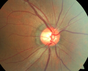
Optic nerve photos

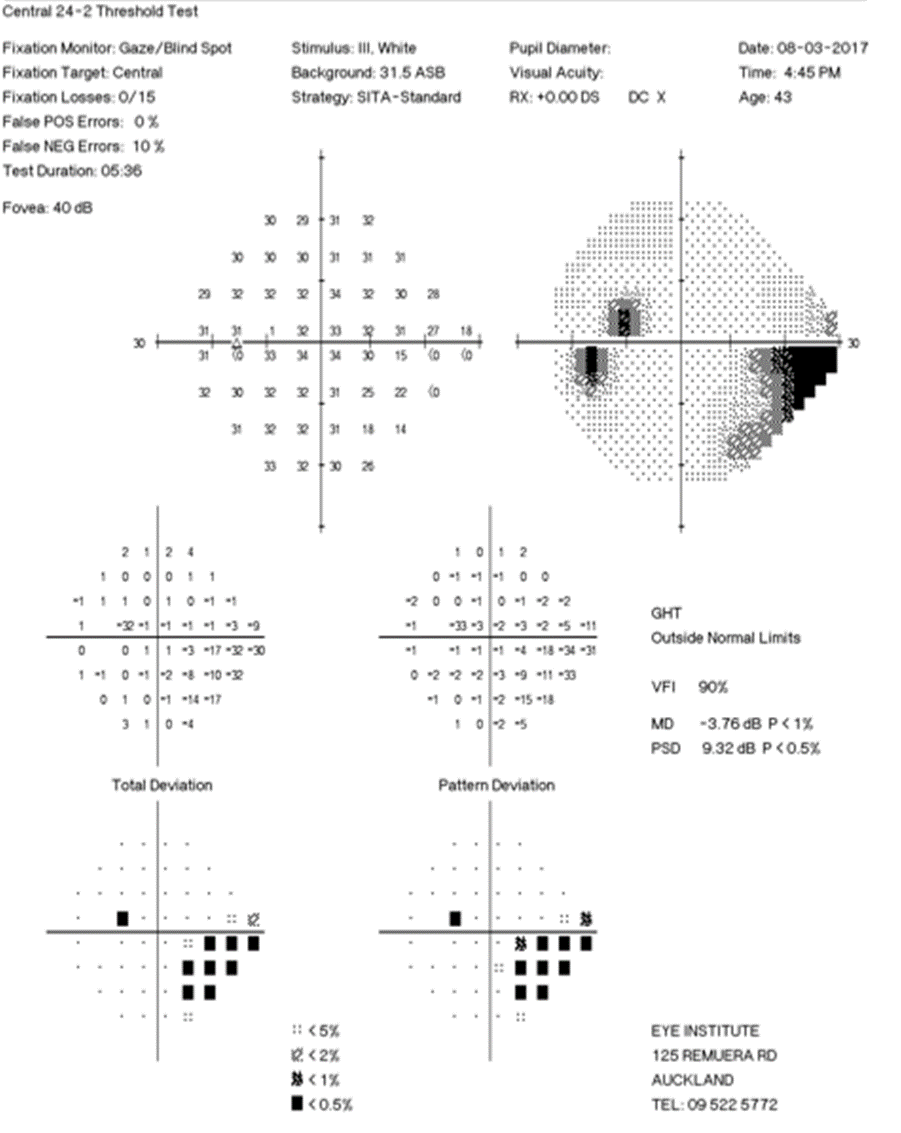
Visual Fields

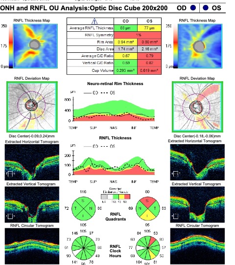
Optical Coherence Tomograph
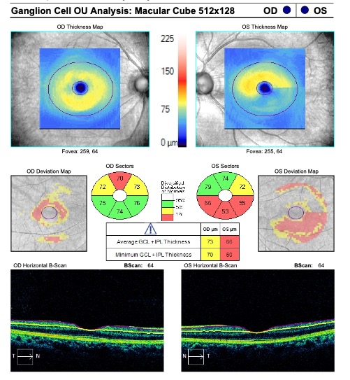
Optical Coherence Tomography – Ganglion Cell Complex
Case Content
0% Complete
0/12 Steps