
Question 5. Describe what are false positives and how it is deternined by automated visual field testing.
Question 5. Describe what are false positives and how it is deternined by automated visual field testing.
Answer:
False positives are recorded when the patient responds without a presented stimulus. False positives up to 33% are acceptable, as per the manufacturer. Excess false positives mask the underlying visual field defects. False positives are generally a mark of unreliable test performance. They are not caused by eye disease or testing artifacts. Rarely, if a patient experiences photopsias it can be cause false positives. Although the perimeter alerts when the false-positive rate is ≥ 33%, fields with a ≥ 5% false-positive rate should be interpreted with caution. Visual fields with ≥ 10% false positives are questionable.
Visual fields can be minimised by patient education. In particular, patients should be informed that it is normal to not seem some of the stimuli. This is a very common reason for false-positives.
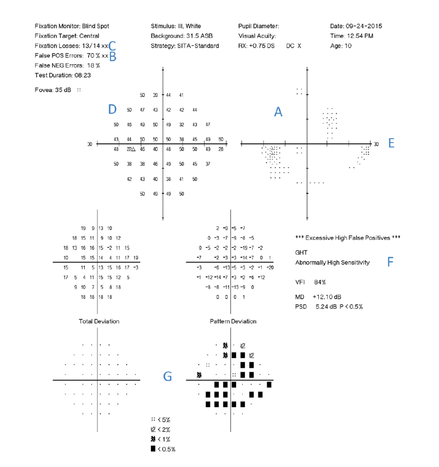
There are seven findings related to an excessively high false-positive rate. A. White scotoma on gray scale. B. High false positives. C. High fixation loss rate. The field analyzer records any response to a stimulus projected onto the physiologic blind spot as a fixation loss, which occurs with increased frequency in an examination with a high false-positive rate. D. Supra-normal sensitivities (higher than foveal sensitivity, which is 35 dB in this case). E. Loss of the physiologic blind spot. F. The Glaucoma Hemifield Test message “Abnormally High Sensitivity,” which appears when the overall sensitivity in the best part of the field is higher than that found in 99.5% of the population. G. “Reverse cataract” pattern in which generalized depression occurs on the pattern deviation rather than total deviation