
Question 4: What is your working diagnosis?
An MRI of the brain is ordered. It shows minor asymmetry in diameter of the optic nerves but no altered signal and no abnormal enhancement. There was no orbital or retro-orbital mass lesion detected.
Question 4: What is your working diagnosis?
Answer
This patient presents with a complex ocular picture.
First, his right eye has a presumed hemi-retinal vein occlusion, previously treated with PRP laser. He now presents with new subretinal and sub-retinal pigment epithelium (RPE) haemorrhages.
Second, his optic discs show evidence suggesting glaucomatous optic neuropathy. The right eye has open angle glaucoma with an old CRVO. The left eye shows no features of glaucoma but given that he is using topical Lumigan, he may have had ocular hypertension , which was presumably well controlled because of the drops.
This case highlights the importance of a thorough history and examination in managing patients with potentially overlapping pathologies. A 2019 meta-analysis assessed the relationship between glaucoma and retinal vein occlusion (RVO) risk with 15 studies meeting the inclusion criteria. The relationship between glaucoma and RVO is somewhat controversial. While analyses, including the Blue Mountains Eye Study, (Cugati et al. 2006) suggest glaucoma as a key risk factor for RVO, others like the Beaver Dam Eye Study (Klein et al. 2000) and Singapore Malay Eye Study (Lim et al. 2008) studies find no significant association after controlling for age.
The patient returns to you 5 years later. His right eye is shown in Figure 4.
Figure 4
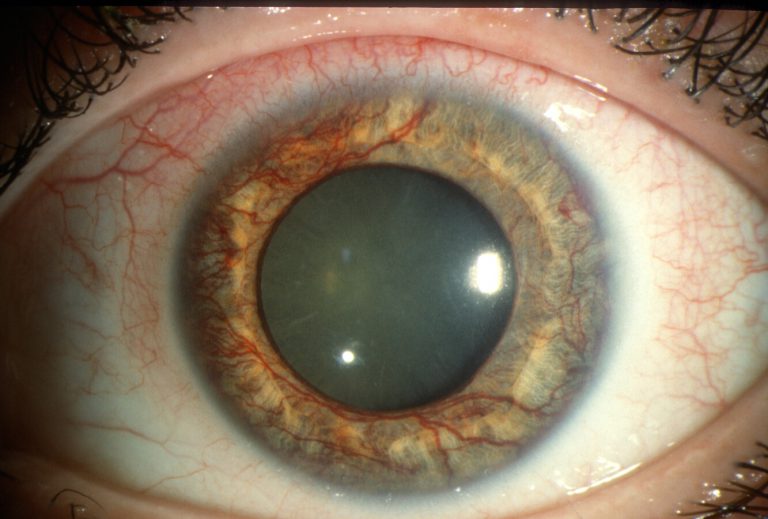
Reproduced from https://morancore.utah.edu/section-10-glaucoma/neovascularization-of-the-iris-rubeosis-iridis/