
Question 1:Describe the colour fundus photographs in Figure 1.
Figure 1: Colour photographs
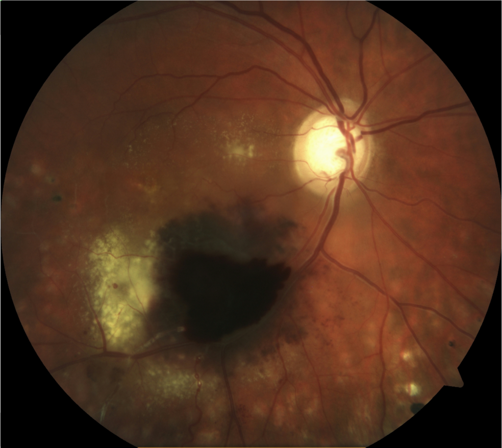
Right eye
Left eye

Question 1:Describe the colour fundus photographs in Figure 1.
Answer:
Both eyes show glaucomatous appearing optic discs.
In the right eye there is collection of subretinal and sub-RPE blood over the temporal arcade, as well as hard exudate. The right eye has a large optic cup measuring 0.85 with increased pallor and superior thinning.
The left eye optic is cupped with a cup to disc ratio is 0.6. The macula and flat and the peripheral retina is unremarkable.
Answer ends