
Case 3 – 2024
Learning Outcomes
- To understand the definition and pathophysiology of non-arteritic anterior ischaemic optic neuropathy (NAION)
- To recognise the clinical presentation of CRVO
- To identify the risk factors associated with CRVO
- To understand the management options for CRVO
History
A 55 year old male reports spots in his vision in his left eye over the last two days. These spots do not move around. He has had treatment in the past for glaucoma
Previous ocular history: Right central haemorrhage with previous laser ~12 years ago
Previous medical history: High cholesterol
Current medications: Lumigan at night, both eyes
Family history: Nil
Examination
Examination findings on presentation shown below.
| Right eye | Left eye | |
| Best corrected visual acuity | 6/60 | 6/4.8 |
| Subjective refraction | -0.25 / 0 x 0 | -1.00 / 0 x 10 |
| Goldmann tonometry | 16 mmHg | 17 mmHg |
| Pachymetry | 499 µm | 499 µm |
| Gonioscopy | Open angles
No evidence of neovascularisation |
Open angles
No evidence of neovascularisation |
| Ishihara | 12/14 (Slow) | 14 / 14 |
| Pupils | R RAPD | |
| Anterior segment | Early lens opacification, otherwise unremarkable | Early lens opacification, otherwise unremarkable |
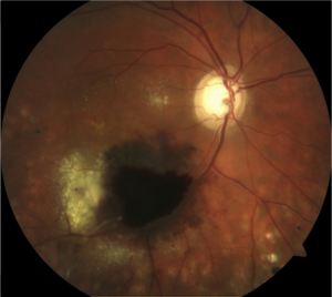
| Fundus examination | See image
PRP over the inferior retina outside of the arcades |
See image |
| Visual fields | See visual fields (Figure 1) | |
| OCT | See RNFL analysis (Figure 2) and GCC scan (Figure 3) | |
Figure 1: Colour photographs
Right eye
Left eye
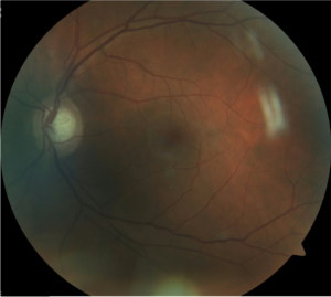
Figure 2: Visual fields
Right eye

Left eye
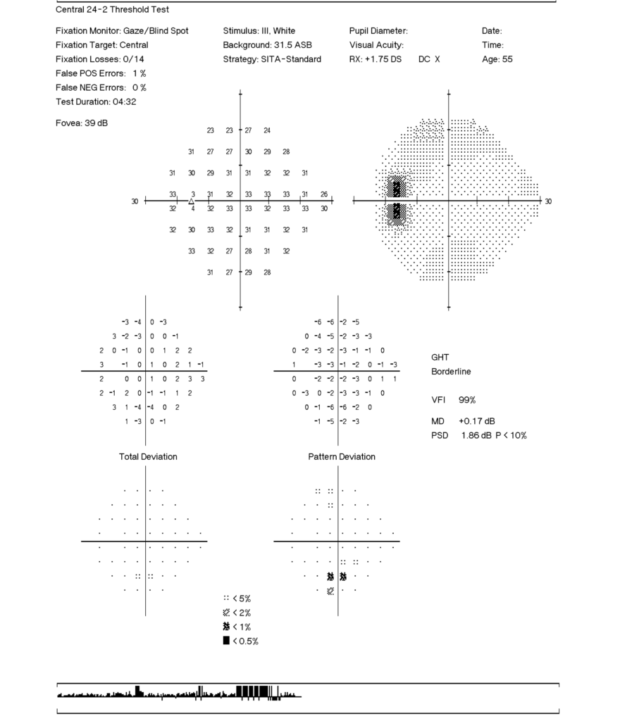
Figure 3: RNFL thickness analysis
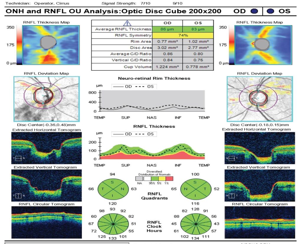
Figure 4
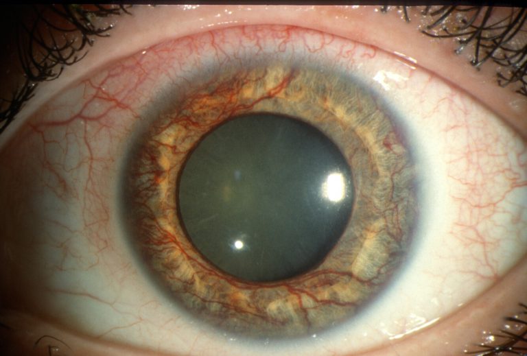
Reproduced from https://morancore.utah.edu/section-10-glaucoma/neovascularization-of-the-iris-rubeosis-iridis/
Detection and prognostic significance of optic disc hemorrhages during the Ocular Hypertension Treatment Study
Association of glaucoma with risk of retinal vein occlusion- A meta-analysis
Anti-vascular endothelial growth factor for neovascular glaucoma
Primary angle closure and primary angle closure glaucoma in retinal vein occlusion
Neovascular glaucoma - A reviewLong-term outcomes of neovascular glaucoma treated with and without intravitreal bevacizumab
Etiology, pathogenesis, and diagnosis of neovascular glaucoma