
Question 5: Describe this patient’s RNFL OCT scans and Ganglion cell layer scan
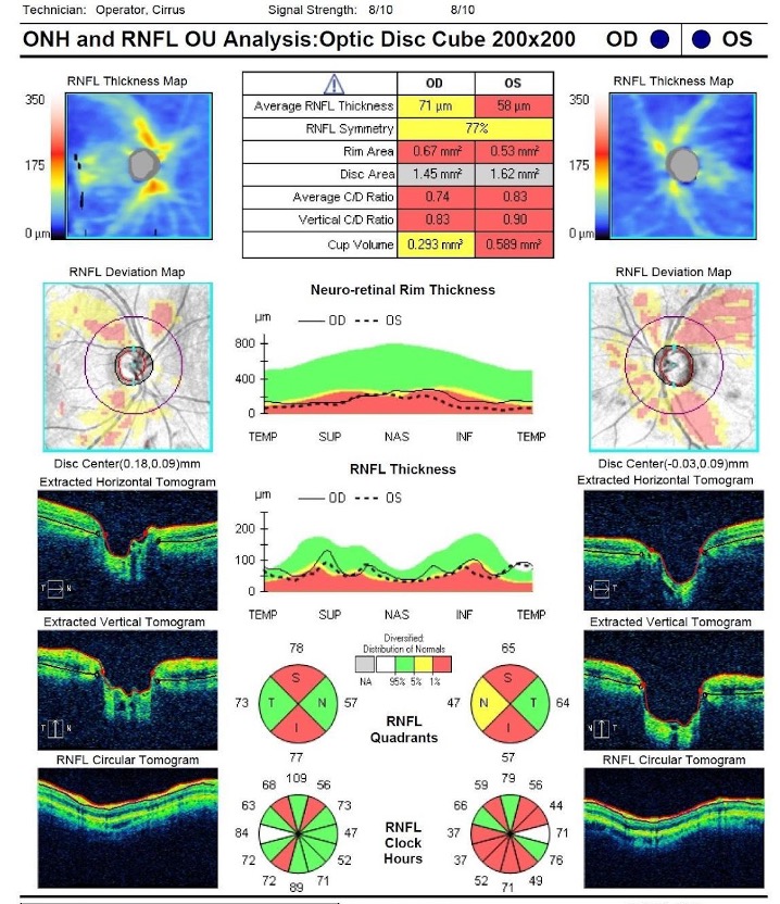
Figure 4: Retinal nerve fibre layer OCT scan analysis both eyes
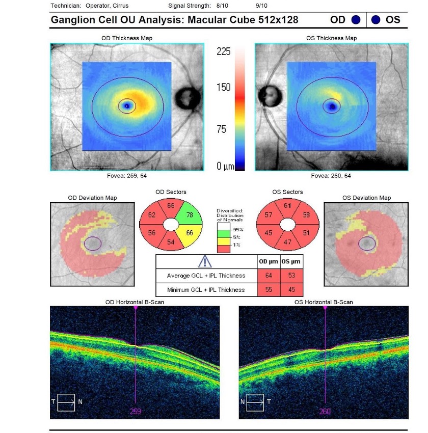
OCT macular scan ganglion cell complex both eyes
Question 5: Describe this patient’s RNFL OCT scans and Ganglion cell layer scan
Answer
The RNFL OCT scans are of adequate quality with no segmentation errors or movement artefacts. The optic disc OCT scan further emphasises the asymmetry between the two eyes. Both eyes show a generalised loss of superior and inferior retinal nerve fibres in both eyes, with more pronounced loss in the left eye, corresponding to the observed thinning of the neuroretinal rim. Additionally, the scan shows that the optic cup is more excavated in the left eye.
The ganglion cell complex (GCC) scan, also of good quality, reveals diffuse thinning of the GCC (including the ganglion cell layer and inner plexiform layer) in both eyes. However, the thinning is more pronounced in the left eye, consistent with the findings on the RNFL OCT and visual field. Furthermore, there is no macular pathology that could confound scan results.
Based on the clinical examination, history, visual field results, and OCT scans, the patient has pigmentary glaucoma, affecting the left eye more than the right eye. The clinical examination and history are consistent with the identified visual field loss and OCT abnormalities. No other causes for visual field loss and OCT findings could be identified.